Нестабильность акромиально-ключичного сочленения
Нестабильность плечевого сустава - это состояние, при котором возникают симптомы выскакивания, "соскальзывания" или выхода плеча из сустава. Обычно она возникает у молодых активных людей, перенесших вывих плеча, но иногда может встречаться и у пожилых людей. Обычно она возникает после травмы плеча ("травматическая нестабильность"), но иногда может возникнуть и при отсутствии травмы ("атравматическая нестабильность"). Это также известно как "рецидивирующий вывих" плеча.
Нестабильность акромиально-ключичного сочленения
Акромиоклавикулярная нестабильность Это заболевание, при котором поражается сустав между акромионом (костным выступом на лопатке) и ключицей (ключичным суставом). Этот сустав необходим для правильного функционирования плечотак как он обеспечивает широкий диапазон движений. Однако, когда связки, поддерживающие этот сустав, растягиваются или разрываются, это может привести к нестабильности и боли.
Нестабильность акромиально-ключичного сочленения может возникнуть по разным причинам, например, из-за травмы, повторяющихся движений над головой или дегенеративных изменений в суставе. Травматические повреждения, такие как падения или прямые удары по плечу, являются наиболее распространенными причинами нестабильности акромиально-ключичного сустава. Эти травмы могут привести к растяжению или разрыву связок, что приводит к нестабильности и боли в плече.
Причины нестабильности акромиально-ключичного сочленения
Существует несколько факторов, которые могут способствовать развитию нестабильности акромиально-ключичного сочленения. Одной из основных причин является травма плеча, например, падение или прямой удар. Эти травмы могут привести к растяжению или разрыву связок, поддерживающих акромиально-ключичный сустав, что приводит к нестабильности.
Повторяющиеся действия над головой, такие как метание или поднятие тяжестей, также могут способствовать разработка нестабильности акромиально-ключичного сочленения. Эти виды деятельности оказывают значительную нагрузку на плечо сустава, что приводит к износ и разрыв связок с течением времени.
Кроме того, дегенеративные изменения в суставе также могут стать причиной нестабильности акромиально-ключичного сочленения. С возрастом хрящ в суставах естественным образом носит В результате они становятся более уязвимыми для травм и нестабильности.
Симптомы нестабильности акромиально-ключичного сочленения
Симптомы нестабильности акромиально-ключичного сочленения могут варьироваться в зависимости от степени тяжести заболевания. К общим симптомам относятся:
- Боль в плече: Боль в плечевом суставе - один из основных симптомов нестабильности акромиально-ключичного сочленения. Боль может быть от слабой до сильной и усиливаться при определенных движениях или действиях.
- Слабость в плече: Нестабильность акромиально-ключичного сустава может привести к слабости плеча. Вы можете заметить снижение способности поднимать или переносить предметы, а также трудности с выполнением действий над головой.
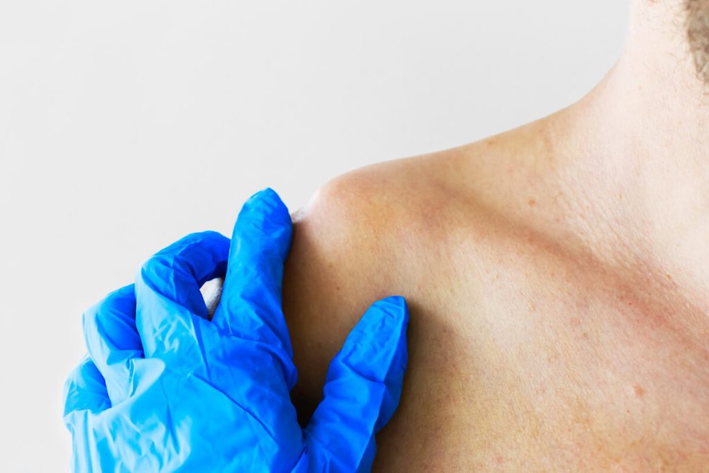
- Отек и нежность: Воспаление и отек вокруг акромиально-ключичного сустава - распространенные симптомы нестабильности. Область может быть нежной на ощупь и выглядеть опухшей или ушибленной.
- Ощущение щелчка или хлопка: У некоторых людей при движении рукой в плечевом суставе может возникать ощущение щелчка или щелчка. Это может быть признаком нестабильности сустава.
Варианты лечения нестабильности акромиально-ключичного сочленения
Варианты лечения нестабильности акромиально-ключичного сочленения зависят от тяжести состояния и конкретных потребностей пациента. В легких случаях может быть достаточно нехирургических методов лечения, в то время как более тяжелые случаи могут потребовать хирургического вмешательства.
Нехирургические методы лечения нестабильности акромиально-ключичного сочленения
Нехирургические методы лечения нестабильности акромиально-ключичного сочленения включают:
- Покой и иммобилизация: Если дать суставу время на заживление и избегать действий, которые усиливают симптомы, это поможет уменьшить боль и способствовать выздоровлению. Для поддержки сустава в процессе восстановления может быть рекомендована иммобилизация, например ношение перевязи.
- Физиотерапия: Физиотерапевт может провести вас через упражнения и растяжки, чтобы улучшить силу и стабильность плечевого сустава. Он также может использовать такие методы, как ультразвук или электростимуляция, чтобы уменьшить боль и воспаление.
Варианты хирургического лечения нестабильности акромиально-ключичного сочленения
Хирургическое вмешательство может потребоваться людям с тяжелой нестабильностью акромиально-ключичного сочленения или тем, кто не реагирует на нехирургические методы лечения. Конкретное хирургическое вмешательство зависит от степени нестабильности и сопутствующих травм.
К распространенным вариантам хирургического вмешательства относятся:
Во время реконструктивной операции поврежденные связки восстанавливаются или заменяются трансплантатами, чтобы вернуть суставу стабильность. Эта процедура обычно выполняется артроскопически, с использованием небольших разрезов и инструмента, наводимого с помощью камеры.
В некоторых случаях может потребоваться стабилизация акромиально-ключичного сустава с помощью винтов, пластин или другого оборудования. Часто это делается в сочетании с реконструкцией связок для обеспечения дополнительной поддержки.
Реабилитация и восстановление после операции по устранению нестабильности акромиально-ключичного сочленения
После операции по поводу нестабильности акромиально-ключичного сочленения необходимо пройти курс реабилитации, чтобы восстановить силы и полноценную функцию плечевого сустава. Процесс реабилитации обычно включает в себя сочетание упражнений, мануальной терапии и постепенного укрепления.
На начальных этапах реабилитации основное внимание уделяется уменьшению боли и воспаления, улучшению диапазона движения и постепенному восстановлению функциональных движений. По мере выздоровления будут включены упражнения для укрепления мышц вокруг плечевого сустава.
Вначале сеансы физиотерапии могут быть назначены несколько раз в неделю, а затем постепенно сокращаются по мере того, как человек набирает силу и стабильность. Важно следовать предписанной программе реабилитации и посещать все запланированные сеансы терапии, чтобы оптимизировать процесс восстановления.
Профилактика нестабильности акромиально-ключичного сочленения
Хотя некоторые случаи нестабильности акромиально-ключичного сочленения являются результатом травматических повреждений, которые трудно предотвратить, есть шаги, которые вы можете предпринять, чтобы снизить риск:
–Укрепляющие упражнения: Регулярное выполнение упражнений, направленных на работу мышц вокруг плечевого сустава, поможет улучшить устойчивость и снизить риск травм.
-Правильная техника: При занятиях, связанных с движениями над головой, например, при метании или поднятии тяжестей, важно использовать правильную технику и избегать чрезмерной нагрузки на плечевой сустав.
–Постепенное развитие: Приступая к новым упражнениям или занятиям, постепенно увеличивайте интенсивность и продолжительность, чтобы дать организму адаптироваться и свести к минимуму риск травм от чрезмерной нагрузки.
–Защитное снаряжение: Если вы занимаетесь контактными видами спорта или деятельностью, связанной с высоким риском получения травмы плеча, подумайте о том, чтобы носить соответствующее защитное снаряжение, например, наплечники или брейсы.
Вывих плеча обычно приводит к повреждению связок плеча, лабрума ("бампера") или костного ободка впадины плеча. Наиболее распространенной травмой является отрыв передней лабумы (так называемое "повреждение Банкарта"). Также может возникнуть вдавливание шара плеча (повреждение Хилла-Сакса). Некоторые люди с "рыхлыми" суставами могут страдать от повторяющейся нестабильности из-за того, что их мышцы работают ненормально (так называемая нестабильность из-за ненормального "мышечного рисунка").
Диагноз "нестабильность плечевого сустава" ставится на основании анамнеза повторяющихся эпизодов выхода плеча из сустава. Иногда пациенты могут испытывать симптомы "мертвой руки" при занятиях спортом или других видах деятельности при отсутствии вывиха. При осмотре могут быть выявлены признаки расшатанности или "рыхлости" в нескольких суставах, боль при определенных движениях плеча и признаки страха или ощущения, что плечо может вывернуться при определенных положениях. Рентген необходим для поиска повреждений костного обода впадины или вмятины на шаре сустава. Для получения дополнительной информации о состоянии лабрума и связок может потребоваться специальная визуализация с помощью магнитно-резонансной томографии. В некоторых случаях может быть запрошена МР-артрограмма (МРТ после введения контрастной жидкости в сустав). Для оценки повреждения костного обода впадины (гленоида) или шара сустава (головки плечевой кости) может быть назначена компьютерная томография.
Риск рецидива травматической нестабильности зависит от пола и возраста, в котором произошел первый эпизод. Риск рецидива ниже у женщин и выше у молодых мужчин в возрасте до 25 лет и снижается с возрастом.
На ранней стадии симптомы можно контролировать с помощью изменения активности.
Физиотерапия под наблюдением врача: Вам может быть рекомендовано обратиться к физиотерапевту, чтобы начать выполнять специальные упражнения для улучшения положения лопаток и укрепления вращательной манжеты.
Сайт Видео Британского общества локтя и плеча (BESS) о нестабильности плечевого сустава содержит полезные рекомендации и упражнения для пациентов с нестабильностью плечевого сустава.
Физиотерапия часто является основным методом лечения пациентов, у которых симптомы нестабильности развились в отсутствие травмы.
Брекеты: Специальный бандаж для плеча может использоваться в течение коротких периодов времени, чтобы защитить плечо и помочь спортивным людям до конца сезона.
Хирургия: Хирургическое вмешательство может быть целесообразным после однократного вывиха у людей с высоким риском рецидива или в тех случаях, когда человек перенес более одного эпизода нестабильности после первой травмы. Это сугубо индивидуальное решение, которое должно приниматься после детального обсуждения со специалистом-хирургом. Операция будет зависеть от патологии, выявленной при клиническом и рентгенологическом обследовании. Она может состоять из артроскопического восстановления, когда разрывы лабрума и связок устраняются с помощью операции "замочная скважина". В случаях, когда имеется значительное повреждение кости обода впадины или шара сустава, могут потребоваться дополнительные процедуры, такие как "ремплиссаж" или костная реконструкция обода впадины.
Более подробную информацию о хирургическом лечении вы найдете в разделе "Процедуры".