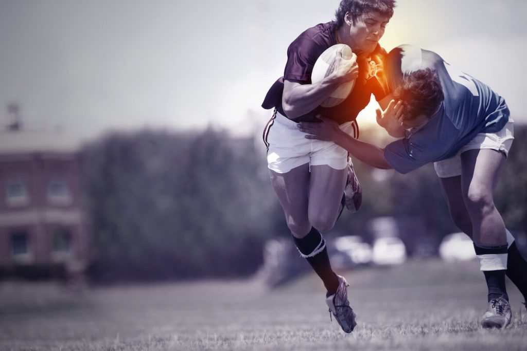
Shoulder Instability
Shoulder Instability is a condition that results in symptoms of popping, “slipping” or the shoulder coming out of joint. It commonly occurs in young active individuals who have suffered a dislocation of the shoulder but may occasionally occur in older individuals. It usually occurs after an injury to the shoulder (“traumatic instability”) but may sometimes occur in the absence of an injury (“atraumatic instability”). It is also known as “Recurrent dislocation” of the shoulder.

Shoulder Instability
Shoulder instability refers to a condition in which the shoulder joint is prone to dislocation or subluxation, where the ball of the upper arm bone (humerus) slips out of the socket (glenoid). This can cause significant pain, limited mobility, and a feeling of insecurity in the shoulder. Understanding the causes, symptoms, and treatment options for shoulder instability is crucial for individuals who experience this condition.
Anatomy of the Shoulder Joint
To comprehend shoulder instability, it is essential to have a basic understanding of the shoulder joint’s anatomy. The shoulder joint is a ball-and-socket joint formed by the humerus (upper arm bone) and the glenoid (a shallow socket in the shoulder blade). The joint is supported by ligaments, tendons, and muscles that provide stability and facilitate movement. The labrum, a ring of cartilage, also plays a vital role in maintaining shoulder stability.
Causes of Shoulder Instability
Shoulder instability can be caused by various factors. One common cause is traumatic injury, such as a fall or a direct blow to the shoulder, which can result in a dislocation or subluxation. Additionally, repetitive overhead activities or sports that involve excessive shoulder movement, like swimming or throwing, can lead to shoulder instability over time. Certain individuals may also be predisposed to shoulder instability due to their unique shoulder anatomy or laxity of the ligaments.
Types of Shoulder Instability
- Dislocation or subluxation: If the spherical head of the shoulder, called the humeral head, shifts from its normal position, it can result in either a complete dislocation or a partial one, referred to as subluxation or humeral subluxation. This is typically associated with acute injuries.
- Labrum Tear: When the labrum, the cartilage responsible for stabilizing the shoulder joint, tears or becomes loose, it can result in a shoulder dislocation. Labral tears may occur due to an acute injury, impact, or repetitive overuse.
- Genetic condition: Shoulder instability may arise from a genetic disorder causing lax ligaments, which raises the likelihood of a weaker or less stable shoulder.
Symptoms and Signs of Shoulder Instability
The symptoms of shoulder instability can vary depending on the severity and type of instability. Common signs include recurrent episodes of shoulder dislocation or subluxation, a sensation of the shoulder “giving way,” pain with certain movements or activities, weakness in the affected shoulder, and a feeling of instability or looseness in the joint. These symptoms can significantly impact daily activities and sports participation, leading to decreased quality of life.
Shoulder instability diagnosis
The diagnosis of shoulder instability involves a comprehensive evaluation. The evaluation may include a detailed review of the medical history, a physical examination to assess range of motion, stability and strength, and imaging tests such as x-rays, MRI or CT scans. These diagnostic tools help identify the type and extent of shoulder instability, guiding the treatment plan.
Treatment Options for Shoulder Instability
The treatment of shoulder instability depends on various factors, including the type and severity of instability, the individual’s activity level, and their goals. Non-surgical and surgical options are available to address shoulder instability and alleviate symptoms.
Non-Surgical Treatment Methods
Non-surgical treatment methods are usually the first line of approach for shoulder instability. These may include:
- Physical Therapy: A structured physical therapy program can help strengthen the muscles around the shoulder joint and improve stability. Therapeutic exercises, stretches, and modalities can be prescribed by a physical therapist to address specific weaknesses or imbalances.
- Bracing and Immobilization: In some cases, a shoulder brace or sling may be recommended to provide support and limit movement while the shoulder heals. Immobilization can help prevent further instability episodes and allow the injured structures to heal.
- Activity Modification: Avoiding activities that exacerbate shoulder instability can be beneficial during the healing process. Modifying techniques or using proper form in sports or daily activities can also help prevent future instability episodes.
Surgical Treatment Options
If non-surgical methods fail to provide relief or in cases of severe instability, surgical intervention may be necessary. The specific surgical procedure will depend on the type and extent of shoulder instability, as well as the individual’s overall health and lifestyle. Common surgical options include:
- Arthroscopic Stabilization: This minimally invasive procedure involves using small incisions and a tiny camera (arthroscope) to repair or tighten the torn or stretched ligaments around the shoulder joint.
- Open Stabilization: In certain cases, open surgery may be required to address extensive ligament damage or bony abnormalities. This procedure involves making a larger incision to directly access the shoulder joint and repair the damaged structures.
- Latarjet Procedure: The Latarjet procedure is a surgical option for individuals with chronic shoulder instability associated with bone loss. It involves transferring a piece of bone from the scapula (shoulder blade) to the glenoid to provide added stability.
Rehabilitation and Recovery
After surgical or non-surgical treatment, a structured rehabilitation program is crucial to restore shoulder strength, range of motion, and stability. Physical therapy plays a vital role in the recovery process. The rehabilitation program typically begins with gentle exercises to regain mobility and gradually progresses to more strenuous activities over time. Compliance with the rehabilitation program and following the healthcare professional’s guidance are essential for optimal recovery.
Prevention and Precautions
While not all instances of shoulder instability can be prevented, certain precautions can help reduce the risk of developing this condition. These include:
- Proper Shoulder Conditioning: Maintaining shoulder strength and flexibility through regular exercise and conditioning can help prevent muscle imbalances and instability.
- Avoiding Overuse: Engaging in repetitive overhead activities should be done with caution. Taking regular breaks, using proper technique, and gradually increasing intensity can help minimize the risk of shoulder instability.
- Wearing Protective Gear: In sports or activities that involve contact or potential trauma to the shoulder, wearing appropriate protective gear, such as shoulder pads or braces, can provide an added layer of support and protection.
A dislocation of the shoulder usually results in damage to the ligaments of the shoulder, the labrum (the “bumper”) or to the bony rim of the socket of the shoulder. The most common injury is a detachment of the anterior labrum (referred to as a “Bankart lesion”). It may also result in indentation of the ball of the shoulder (a “Hill-Sachs” lesion). Some individuals with “loose” joints may suffer recurrent instability due to their muscles working in abnormal fashion (referred to as instability due to abnormal “muscle patterning”).
A diagnosis of Shoulder instability is made based on the history of repeated episodes of the shoulder coming out of joint. Occasionally patients may experience symptoms of a “dead arm” with sporting or other activities in the absence of dislocation. Examination may show signs of laxity or “looseness” in multiple joints, pain with certain movements of the shoulder and signs of apprehension or a feeling that the shoulder may come out when placed in certain positions. X-rays are essential to look for damage to the bony rim of the socket or indentation of the ball of the joint. Special imaging with an MRI scan may be requested to obtain further information about the state of the labrum and the ligaments. In some instances an MR arthrogram (MRI scan after injection of contrast fluid in the joint) may be requested. A CT scan may be arranged to assess the damage to the bony rim of the socket (glenoid) or the ball of the joint (humeral head).
The risk of recurrence of traumatic instability depends on gender and the age at which the first episode occurred. The risk of recurrence is lower in women and is greatest in young men below 25 years of age and diminishes with age.
In the early phase, symptoms may be controlled with activity modification.
Supervised physiotherapy: You may be advised to see a physiotherapist to start a regime of specific exercises to improve scapular positioning and strengthen the rotator cuff.
The British Elbow and Shoulder Society (BESS) video on shoulder instability has useful guidance and exercises for patients with shoulder instability.
Physiotherapy is often the main treatment for patients who have developed symptoms of instability in the absence of an injury.
Braces: A specific brace for the shoulder may be used for short periods of time to protect the shoulder and get sporting individuals through to the end of a season.
Surgery: Surgery may be appropriate following a single dislocation in individuals who are at a high risk of recurrence or where an individual has suffered more than one episode of instability following an initial injury. This is a highly individualised decision and should be made after detailed discussion with a specialist surgeon. The operation performed will depend on the pathology identified on clinical and radiological assessment. It may consist of an Arthroscopic repair where the tears in the labrum and ligaments are repaired with “keyhole” surgery. In cases where there is significant bone damage to the rim of the socket or the ball of the joint, additional procedures such as a “remplissage” or bony reconstruction of the rim of the socket may be necessary.
For further information on surgical treatment, please refer to the procedures section.
